
How does the “body” take shape?
The body of animals is highly complex, containing distinct regions that carry out specific functions. As the body forms during embryogenesis, three germ layers are created; these are the endoderm (precursor of the epidermis and neural tissue), mesoderm (precursor of muscles, skeleton, cardiovascular and urogenital tissues) and the ectoderm (precursor of digestive tract and organs). During the process of gastrulation, the germ layers are formed and then rearranged, which creates the internal and external parts of the developing embryo.
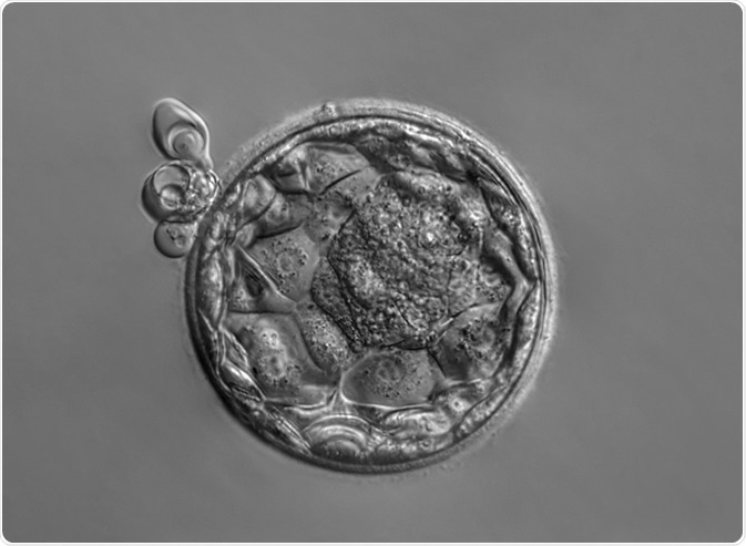 Image Credits: Vladimir Staykov / Shutterstock.com
What processes drive gastrulation?
Gastrulation occurs according to a conserved series of movements defined by their morphogenetic outcome; emboly (internalization), epiboly, and convergence and extension.
Emboly
Emboly, also known as internalization, is the first process in gastrulation. During this process, cells of the blastula which will become the endoderm and mesoderm are moved beneath the layer of cells which will become the ectoderm.
When emboly begins, the progenitor endodermal and mesodermal cells are found in the epithelial layer of the blastula, therefore these cells need to undergo epithelial to mesenchymal transition; here, the junctions of the epithelial cells are broken down, the expression of adhesion proteins are reduced, an intermediate filament network is formed and the microtubule network is rearranged so that it clusters around the centrosome.
Emboly occurs through a structure called the blastopore, and when the progenitor endodermal and mesodermal cells have passed through the blastopore the cells migrate away from the blastopore to form the three germ layers by rearranging beneath the ectoderm layer.
The activities of the transcription factors Snail and Sox3 keep the ectodermal progenitor cells in the epiblast while allowing the migration of mesendodermal progenitor cells through the blastopore.
There are various ways in which emboly occurs, for example, invagination, involution, and ingression.
- Invagination; here, a fold is created in the epithelial cells by the mesoderm folding inwards to form the blastopore, which then takes the mesoderm further inwards as the fold deepens. This type of emboly is seen in the fruit fly Drosophila melanogaster.
- Involution; here, the progenitor mesoderm and the endoderm cells cluster as a tissue around the blastopore. Constriction of so-called bottle cells creates the blastopore opening, and the clustered tissue roll through the blastopore. This type of emboly is seen in frogs.
- What makes ingression distinct from the other types of emboly is that epithelial to mesenchymal transition occurs before these cells are internalized. Here, the progenitor mesoderm and endoderm cells differentiate from the epithelial cells, then move through the blastopore and continue migration as individual cells. This type of emboly is seen in mice.
Epiboly
Epiboly is where the cells of the embryo expand, and this process starts prior to the distinct formation of the three germ layers. During this process, embryos formed from one epithelium (such as mammalian embryos) expand while maintaining the same thickness while embryos formed from multilayered epithelium typically become thinner as it expands. In frog embryo development, epiboly occurs by intercalation of cells forming the deeper layer to the superficial layer, which occurs radially. As this process is not guided by axes of the embryo, epiboly occurs in all directions.
Convergence and extension
During convergence and extension, the germ layers are elongated along the head-tail (anteroposterior) axis while being narrowed along the front-back (dorsoventral) axis. Neural cells and mesendodermal cells that will form the head are positioned close to the cells of the trunk, and during convergence and extension, these cells separate. There are various types of convergence and extension;
- “Convergent extension”; here, mesodermal cells change shape so that they elongate along the mediolateral embryonic axis. Intercalation then occurs between cells that are either medial or lateral, thereby narrowing mediolaterally and elongating at the same time. This was found to occur in frog embryo development.
- Similar effects to convergent extension are possible by either polarized radial intercalation (where cells from different layers connect to another layer) or polarized cell division, where the daughter cells are aligned to a specific embryonic axis.
- Cell migration; it is possible to direct cells to specific areas of the embryo to achieve convergence and extension. For example, in zebrafish migration of the lateral mesoderm is directed to the dorsal side towards the dorsal midline, but the cells which are at the anterior or posterior ends are directed further towards the anteroposterior axis.
Sources
Solnica-Krezel, L. and Sepich, D. S. (2012) Gastrulation: Making and Shaping Germ Layers. Annual Review of Cell and Developmental Biology www.annualreviews.org/doi/full/10.1146/annurev-cellbio-092910-154043
Solnica-Krezel, L. (2005) Conserved Patterns of Cell Movements during Vertebrate Gastrulation. Current Biology www.cell.com/…/S0960-9822(05)00277-0
Maroto, M. et al. (2012) Somitogenesis. Development https://dev.biologists.org/content/139/14/2453
Last Updated: Mar 12, 2020

Written by
Dr. Maho Yokoyama
Dr. Maho Yokoyama is a researcher and science writer. She was awarded her Ph.D. from the University of Bath, UK, following a thesis in the field of Microbiology, where she applied functional genomics toStaphylococcus aureus . During her doctoral studies, Maho collaborated with other academics on several papers and even published some of her own work in peer-reviewed scientific journals. She also presented her work at academic conferences around the world.
Source: Read Full Article