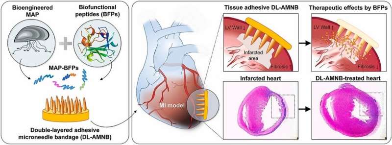
Patients not only have to fight their diseases but also must deal with the pain—the more severe the condition, the more painful the injections. Recently, new doors have opened to relieve patients of these burdens. A POSTECH research team has developed a treatment for myocardial infarction using a MAP-based microneedle bandage that are attachable to the heart tissue.
A research team led by Professor Hyung Joon Cha and Ph.D. candidate Soomee Lim, Dr. Tae Yoon Park, and Dr. Eun Young Jeon (currently at Columbia University) of the Department of Chemical Engineering at POSTECH has developed new mussel adhesive proteins (MAPs) that incorporate biofunctional peptides derived from growth factors or extracellular matrix in the body. Growth factors are proteins involved in cell growth and differentiation, and extracellular matrix refers to the rest of the tissue excluding the cells.
The research team constructed a microneedle, a patch-type injection that is attached to the heart tissue to effectively deliver the biofunctional peptide to the damaged myocardial tissue. Silk fibroin protein, which displays excellent mechanical strength, was added to the tip of the microneedle to facilitate and accelerate its penetration to the surface of the myocardial tissue in the animal model.
Microneedles made up of micro-sized needles of 300 to 800 micrometers (㎛) long have been investigated as a drug delivery system that delivers active ingredients through microchannels. Unlike conventional injections which require a thick hypodermic needle, the microneedle can relieve patients’ pain and allow easy application to the surface of the organ tissue.
The research team applied this microneedle platform to the treatment of myocardial infarction. When myocardial infarction occurs, the cardiac muscle cells and surrounding blood vessels are severely damaged, but no method existed to regenerate cardiac muscles since they did not regenerate themselves. Though research is being conducted to regenerate damaged tissues by delivering growth factors or therapeutics to the heart for blood vessel regeneration, growth factors have a very short half-life and rapidly clear in the body, requiring continuous injections.
In this study, the researchers demonstrated that when human-derived vascular cells were treated with MAPs containing biofunctional peptides, cell proliferation and migration were effectively promoted. Thanks to MAP’s excellent adhesiveness and swelling behavior of the microneedle, the patch was firmly retained in the heart tissue that has continuous, repetitive contraction movements.
The functional MAPs were delivered directly through a microscopic pathway created by the microneedle and remained in the damaged myocardial tissue for a prolonged period, preventing further death of myocardial cells and effectively restoring the damaged myocardial wall by alleviating fibrosis.
“We have effectively delivered biofunctional peptides in an animal myocardial infarction using a mussel adhesive protein, a biomaterial that originated in Korea,” explained Professor Hyung Joon Cha. “This not only confirms the potentiality of the newly developed treatment for myocardial infarction, but also of its applicability to tissue regeneration treatment in similar environments.”
Source: Read Full Article
