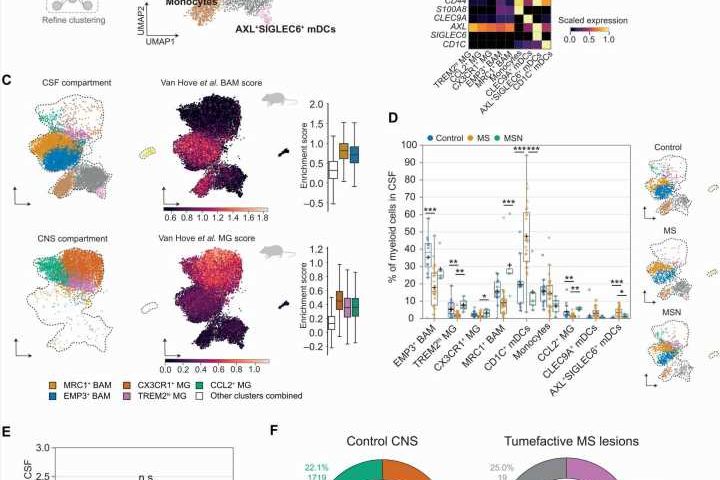
 Subsetting strategy and manifold plot for the myeloid lineage. (B) The matrix plot shows a selection of marker genes for the respective clusters. (C) Manifold plots show the dataset subsetted to either CSF (top left) or CNS samples (top right) and the expression of murine BAM-specific (bottom left) or MG-specific (bottom right) transcription profiles; box plots indicate profile expression between clusters (27). (D) The box plot shows the frequencies of myeloid subsets in CSF for control (n = 12), MS (n = 24), and MSN (n = 5) (left) with condition-specific manifold plots downsampled to show equal number of cells for each condition (right). Statistics were computed using linear regression. (E) Flow cytometry cell counts (in cells/ml) are shown for mDCs in CSF of control (n = 7), MS (n = 25), and MSN (n = 4). (F) Donut charts of CNS samples are shown for either control (n = 9) or lesional tumefactive MS tissue (n = 3) with integrated manifold plots. Box plots show the IQR with whiskers indicating 1.5 × IQR, “+” indicating the mean, and the line indicating the median. *P Science Translational Medicine (2022). DOI: 10.1126/scitranslmed.adc9778")
For diagnosis of and therapy decisions on neurological diseases, the cerebrospinal fluid is often key. The cells in this fluid may also provide information on pathological processes that occur in the brain. Of course, this requires distinguishing between cells derived from the brain and cells that stem from the blood. Until now, this was hardly possible.
Neuroscientists from the University of Münster have now generated an atlas that could change this—using it will allow a better understanding of disease mechanisms and a new way to study the effects of therapies. This invaluable resource is provided as a freely available software for researchers all around the world.
Imagine a traffic jam on the highway, and to bypass it, drivers would be recommended to use a hiking map. Of course, the provided routes would be useless. For a long time, a similar problem was faced in the field of neuroscience. Even though immune cells were found in the brain and cerebrospinal fluid (CSF) of patients, scientists had to rely on atlases based on peripheral blood to characterize the phenotype and origin of a cell.
As a consequence, the navigation of the central nervous system (CNS) was erroneous and predictions were often wrong. The latest work of scientists from the Westphalian Wilhelms-University Muenster (WWU) corrects this by proving an atlas of CNS- and CSF immune cells. Their findings were recently published in Science Translational Medicine.
The exact classification of cells and their tissue of origin is necessary for a better understanding of diseases and their progression. The new atlas not only provides a detailed characterization of a CSF immune cell’s identity, but also predicts where it comes from—the blood or the CNS.
To generate the atlas, researchers from the working group of Prof. Nicholas Schwab at the University Hospital of Muenster used cutting-edge technology that analyzes the gene expression of singular cells. Using so-called deep learning algorithms, the information was then combined with large datasets from the neurology department in Muenster and other international research institutions. Using this approach, the results were generalizable. The scientists, especially Dr. Patrick Ostkamp, combined data from both the CNS and the CSF.
A primary mechanism of MS pathology is the migration of peripheral blood immune cells into the CNS, where they eventually cause damage. “Cells never act without a reason. Therefore, it is very important to know whether a cell is heading from the blood to the CNS, or whether it has been there already and is on its way back,” says Prof. Nicholas Schwab, coordinator of the study.
To examine this, the team used CSF from patients that were treated with a drug that blocks the entry of cells into the CNS. The CSF of these patients can be regarded as a one-way street and most cells that were found in the CSF must have been derived from the CNS, as entry from the blood was blocked. Indeed, the CSF of these patients not only harbored fewer cells but the composition of immune cells was skewed as well. Comparison to cells obtained through CNS biopsies confirmed that the CSF now mostly contained cells that were alike to CNS cells.
In addition, Parkinson’s and Alzheimer’s disease samples were included as well, since recent studies indicated that the immune system may play a pivotal role in these diseases as well. Therefore, the new atlas from Muenster may also shed light on the immunological landscape of other neurological diseases.
To provide this useful tool to other researchers, the group developed a software to help the international research community navigate through the complexity of the immune system of the CNS. The scientists hope that in the long run their results will not only assist researchers in their scientific endeavors, but that they may also be used to help patients.
Prof. Heinz Wiendl, head of department of neurology in Muenster says, “This innovative project confirms what neurologists have assumed for more than a century: the CSF is a mirror of the CNS. It will help to interpret what the CSF indicates and this will be of enormous value in clinical practice.”
Importantly, the CSF—in contrast to the CNS—is attainable via lumbar puncture. “Data from the CSF can therefore shed new light on molecular mechanisms of drugs and immunological differences between diseases. The analysis of the CSF can therefore be considered a ‘CNS biopsy light.’ This information is invaluable.”
More information:
Patrick Ostkamp et al, A single-cell analysis framework allows for characterization of CSF leukocytes and their tissue of origin in multiple sclerosis, Science Translational Medicine (2022). DOI: 10.1126/scitranslmed.adc9778
Journal information:
Science Translational Medicine
Source: Read Full Article