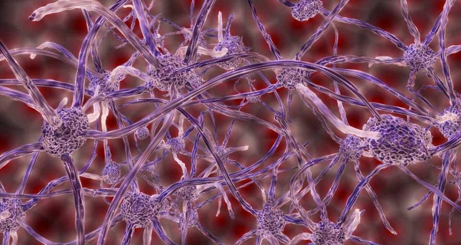
In a study published in Nature Neuroscience, scientists at the Center for Excellence in Brain Science and Intelligence Technology (CEBSIT) of the Chinese Academy of Sciences, along with their collaborators, reported the first release of a whole-brain projectome comprising over 6,000 single neurons in the mouse prefrontal cortex (PFC), making it the largest database of a whole-brain, single-neuron mouse projectome to date.
The scientists identified 64 projectome-defined neuron subtypes in the mouse PFC and their spatial organization, the modularity and hierarchy of intra-PFC connectivity, and the correspondence between transcriptome-defined and projectome-defined neuron subtypes.
The information flow between different brain regions in the cerebral cortex relies on the long-range axonal projections of neurons. Neurons with different projection patterns are often involved in distinct brain functions.
Therefore, studying the projection patterns of individual neurons is crucial for understanding organization and information processing in the brain. Reconstructing the whole-brain projectome at the single-neuron level can identify new neuron subtypes and uncover wiring rules of the brain network, thus allowing more systematic understanding of how the brain works.
Currently, scientists around the world are working intensively to study the whole-brain projectome at the single-neuron level.
In the brain, the PFC is the center of high-level cognitive function including decision-making, working memory, and attention. Abnormality and malfunction of the PFC can cause many neuropsychiatric diseases. Axonal projections of the PFC cover nearly all brain areas including the cortex, striatum, thalamus, midbrain, and hindbrain. The axonal projection patterns of neurons in the PFC at the single-cell level are still unclear and the modular organization of the connectivity network within the PFC remains to be uncovered.
Reconstructing the whole-brain projectome at single-cell resolution in mammalian brains is a daunting task and requires continuous tracing of single neurons one-by-one, using large-scale, TB-sized, light microscopic imaging data in 3D. The entire tracing process is labor-intensive, extremely complex, and time-consuming.
To solve this problem, Dr. Gou Lingfeng, a researcher at Dr. Yan Jun’s lab, developed the software package Fast Neurite Tracer (FNT). It facilitated large-scale study of the single-neuron projectome by enabling high-throughput, single-neuron projectome reconstruction and analysis of TB-sized, light microscopic imaging data.
Collaborating with Dr. Xu Ning-long’s lab at CEBSIT, Dr. GONG Hui’s lab at Huazhong University of Science and Technology and many labs at CEBSIT, the researchers reconstructed the complete axon morphology of a total of 6,357 single PFC neurons from the optical imaging data of 161 mouse brains and identified 64 projectome-defined subtypes after quantifying the similarity of axon morphology of different neurons.
Researchers then mapped the spatial distribution of these neuron subtypes in different prefrontal subregions and cortical layers. They analyzed the intra-PFC connectivity network and constructed a high-resolution intra-PFC network, revealing the modular and hierarchical structure within the PFC.
Researchers also performed comparative analysis between transcriptome-defined and projectome-defined neuron subtypes. Using retrograde tracing and single-molecule fluorescence in situ hybridization, they found that each transcriptome-defined subtype corresponds to multiple projectome-defined subtypes.
Source: Read Full Article
