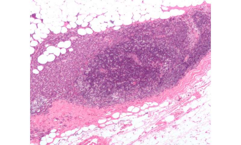
Mammograms are commonly used to screen for breast cancer. In spite of easy access, conventional mammograms cannot find every tumor due to the limited image contrast mechanism.
The measurement of X-ray beam refraction in breast tissues has the potential to be the next generation screening technique for breast cancer. A new technique called X-ray phase contrast imaging (XPCI) provides better soft tissue differentiation and tumor detections.
However, the use of X-ray interferometry made from gold and silicon gratings sharply reduces the X-ray dose efficiency, i.e., inhibiting the patient radiation dose.
Recently, researchers from the Shenzhen Institutes of Advanced Technology (SIAT) of the Chinese Academy of Sciences developed a novel XPCI signal extraction technique using a deep learning method. The technique has shown promising advantages in enhancing signal accuracy and improving the X-ray radiation dose efficiency.
The study was published in IEEE Transactions on Biomedical Engineering on July 22.
The researchers designed a deep convolutional neural network named XP-NET, using a special architecture to automatically perform the XPCI signal retrieval and image quality enhancement in a sequence.
Results showed that the XP-NET was able to improve the phase signal accuracy by over 15% compared with the conventional analytical method.
Additionally, both biological specimen and breast phantom studies demonstrated that the phase images acquired with half the radiation dose and processed by the XP-NET showed image quality comparable to the reference images acquired with the standard radiation dose level.
Source: Read Full Article
