A Chopin or Mozart composition loses its meaning and magic when played on a piano that’s out of tune.
And when a person who is profoundly deaf has a cochlear implant, which simulates the normal hearing process by electrically stimulating nerves in the cochlea, the device also needs to be tuned, like the strings on a piano, to achieve perfect pitch.
A team of researchers from Western University, in collaboration with Uppsala University Hospital in Sweden, has designed a mathematical tool to help do just that—program the implant with precision for each patient’s unique anatomy.
The findings are published in the journal IEEE Transactions on Biomedical Engineering.
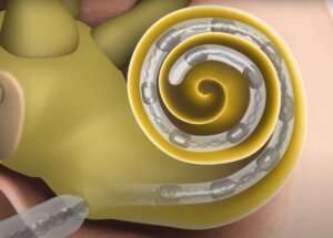
When cochlear implants are surgically placed, their electrodes are inserted along various points of a patient’s cochlea, the spiral-shaped part of the inner ear involved in converting vibrations into pitches of sound.
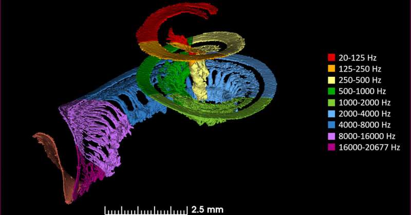
Each part of the cochlea is responsible for a different pitch—a C note stimulates a different part of the spiral than a B-flat note for example. Because each person’s cochlea is a slightly different in size and shape, this distribution varies among individuals.
“Until now, cochlear implant programming was performed in a sort of one-size-fits-all approach,” said Luke Helpard, Ph.D. candidate in the School of Biomedical Engineering. “We’re using imaging data to be able to customize the pitch map for each patient based on individual patient anatomy and post-operative electrode locations within the cochlea.”
A pitch map refers to how stimulation frequencies are assigned to each electrode. If the implant is stimulating with the wrong pitch, it can degrade speech perception as well as dampen the person’s ability to fully appreciate music or nature sounds. This can also increase the amount of time it takes for patients to adapt to an implant if they are working to overcome a pitch mismatch.

The new tool uses measurements of patient-specific anatomy obtained from clinical CT scans. These are input into a mathematical formula developed by the team that calculates exactly how each electrode on the implant should be programmed.
“Typically, when we talk about math, people have a glazed look because they can’t see how it is applicable in real life,” said Hanif Ladak, professor at Western’s Schulich School of Medicine & Dentistry and Faculty of Engineering. “What’s beautiful about this is that our mathematical tool has an obvious real-life application and audiologists are already beginning to evaluate it in patients.”
Ladak is leading the project alongside Dr. Sumit Agrawal, associate professor at Schulich Medicine & Dentistry, and their graduate students Helpard and postdoctoral fellow Alireza Rohani.
Rohani said the team designed the tool based on information gleaned from doing very high-resolution 3D visualizations of the cochlea from cadaveric samples. The imaging was done in collaboration with Canadian Light Source in Saskatoon, a centre that provides a specialized kind of imaging using synchrotron light.
“Imaging the cochlea is very difficult because it is tiny and encased by the densest bone in the body, and we need to be able to visualize both soft tissue and bone,” said Rohani. “This collaboration allowed us to compare the clinical CT to the high-resolution 3D images from Canadian Light Source so we could refine our clinical measurements.”
The World Health Organization estimates that 466 million people around the world have disabling hearing loss.
“Cochlear implants have been a revolutionary advance for these patients,” said Agrawal, a cochlear implant surgeon at London Health Sciences Centre. “This research has the potential to significantly improve their quality of life through music appreciation, understanding tonal languages and comprehending speech in challenging listening environments.”
Source: Read Full Article
