 *Important notice: medRxiv publishes preliminary scientific reports that are not peer-reviewed and, therefore, should not be regarded as conclusive, guide clinical practice/health-related behavior, or treated as established information.
In a recent study posted to the medRxiv* preprint server, researchers at the University of North Carolina and the National Institutes of Health determined the associations between oral severe acute respiratory syndrome coronavirus 2 (SARS-CoV-2), oral antibodies against SARS-CoV-2, and coronavirus disease 2019 (COVID-19) symptoms.
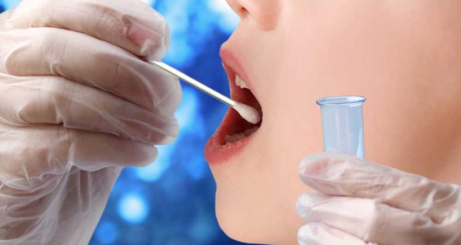
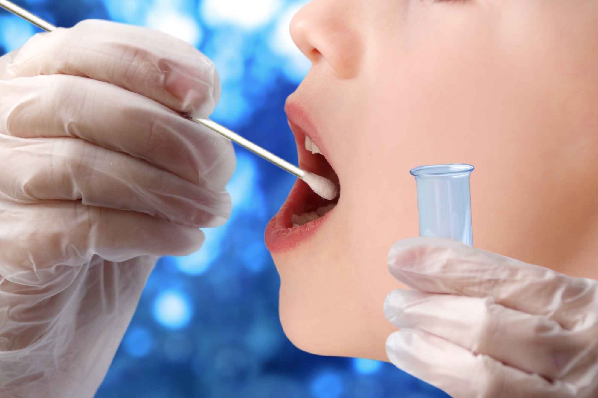 Study: Oral SARS-CoV-2 host responses predict the early COVID-19 disease course. Image Credit: Kittyfly / Shutterstock
Background
SARS-CoV-2, the causative agent of COVID-19, replicates in the upper respiratory tract, oral mucosa, salivary glands, and respiratory mucosa. The presence of angiotensin-converting enzyme 2 (ACE2) receptors and the detection of SARS-CoV-2 ribonucleic acid (RNA) and virulent SARS-CoV-2 in the oral cavity indicates that SARS-CoV-2 proliferates in the oral cavity. While lateral flow assay (LFA)-determined anti-SARS-CoV-2 responses in the oral cavity denote systemic immunity, oral biomarkers as indicators of COVID-19 prognosis have not been explored significantly.
About the study
In the present study, researchers assessed SARS-CoV-2 detection and humoral immune responses of the host in the oral cavity.
Throat wash and saliva samples were obtained from 47 symptomatic (n=17) and asymptomatic (n=30) individuals, for whom COVID-19 diagnosis was confirmed using quantitative reverse-transcription polymerase chain reaction (RT-qPCR) by analyzing nasopharyngeal (NP) swabs. In the asymptomatic group, 15 individuals showed SARS-CoV-2 seropositivity, and the remaining were seronegative or uninfected. SARS-CoV-2 nucleocapsid (N) protein was detected using immunoblot assays.
Quantitative reverse-transcription-polymerase chain reaction targeting SARS-CoV-2 subgenomic ribonucleic acid (sgRNA) sequences were confirmed by Sanger sequencing, and LFA was performed to determine anti-SARS-CoV-2 spike (S) protein receptor-binding domain (RBD) immunoglobulin G (IgG) and IgM titers. In addition, structural analysis was performed to identify molecules in the host's saliva similar to the SARS-CoV-2 nucleocapsid antigen.
COVID-19 severity was classified using the national institute of health (NIH) coronavirus disease 2019 treatment guidelines. Complete-length subgenomic polymerase chain reaction products coding for SARS-CoV-2 spike, nucleocapsid, envelope (E), or membrane (M) glycoproteins were generated from SARS-CoV-2-positive total ribonucleic acid content in saliva. Complementary deoxyribonucleic acid (cDNA) was transfected to human normal oral keratinocytes (NOK), and 48 hours post-transfection, immunoblot analysis was performed.
The crystal structure of the SARS-CoV-2 nucleocapsid antigen N-terminal ribonucleic acid-binding domain was comparatively assessed with publicly accessible structures uploaded in the molecular modeling database (MMDB). Oral cavity samples comprising greater than 10.0 RNA copies per RT-qPCR reaction were considered SARS-CoV-2-positive. COVID-19 symptoms, including muscle pain, weakness, anosmia, nausea, ageusia, upper respiratory tract symptoms, breathlessness, cough, nasal congestion, sore throat, and discharge from the nasal cavity, were assessed.
Results
Antibodies eBook

The average age of the study participants was 40 years, with even gender distribution. At study initiation, immunoblotting analysis confirmed LFA-detected SARS-CoV-2 N antigen presence in 82.0% of the throat washes. However, only three and 17 saliva samples and throat washes, respectively, were SARS-CoV-2-positive by RT-PCR. After four weeks, 60.0% and 83.0% of saliva samples and throat washes, respectively, showed persistent SARS-CoV-2 nucleocapsid antigen presence.
SARS-CoV-2 nucleocapsid antigen lateral flow assay signal among three SARS-CoV-2-negative individuals indicated probable cross-identification of four structurally similar salivary ribonucleic acid-binding protein molecules [alignment 19 to 29 amino acid, root mean square deviation (RMSD) 1.0 to 1.5 Å]. At study initiation, symptomatic patients showed proliferation-related subgenomic ribonucleic acid junctions, and IgG titers (94% and 100% of saliva samples and throat washes, respectively) and IgM titers (75% and 63% of saliva samples and throat washes, respectively).
At four weeks, anti-SARS-CoV-2 immunoglobulin G titers persisted in 100% of saliva samples and 83% of throat washes, and anti-IgM titers persisted in 80% of saliva samples and 67% of throat washes. Oral anti-SARS-CoV-2 IgG titers showed a 100% correlation with the nasopharyngeal swab-analyzed RT-qPCR results. The severity of fatigue and cough and the presence of weakness, nausea, and upper respiratory tract symptoms were inversely related to oral immunoglobulin IgM anti-SARS-CoV-2 titers, which were more significant among women than men. Longitudinal evaluation of symptomatic COVID-19 patients indicated oral SARS-CoV-2 persistence. COVID-19 symptoms and severity correlated with oral anti-SARS-CoV-2 antibody titers and SARS-CoV-2 presence.
The findings provide new insights into the oral biomarkers of COVID-19 prognosis, SARS-CoV-2 transmission and persistence. SARS-CoV-2 presence was detected in oral fluids from nasopharyngeal swab-analyzed RT-qPCR-positive individuals using multiple RT-qPCR-based methods of detection including (i) three distinctive pairs of primers targeting the spike-, open-reading frame 3a (ORF3a)-, or nucleocapsid-coding regions of the SARS-CoV-2 genome; (ii) ribonucleic acid copy numbers in absolute terms; and (iii) subgenomic ribonucleic acid, a biomarker of active SARS-CoV-2 replication in the initial period of symptomatic SARS-CoV-2 infection.
Overall, the study findings showed that critical for SARS-CoV-2 transmission and the course of COVID-19, SARS-CoV-2 proliferation and persistence in the oral cavity demonstrated clear associations with particular COVID-19 symptoms, early immunoglobulin titers, and participant gender during initial infection. Nucleocapsid antigen cross-reactivity might represent mimicry of structurally similar host cell proteins.
*Important notice: medRxiv publishes preliminary scientific reports that are not peer-reviewed and, therefore, should not be regarded as conclusive, guide clinical practice/health-related behavior, or treated as established information.
- Preliminary scientific report. Oral SARS-CoV-2 host responses predict the early COVID-19 disease course, DOI: https://doi.org/10.1101/2023.03.06.23286853, https://www.medrxiv.org/content/10.1101/2023.03.06.23286853v1
Posted in: Medical Research News | Disease/Infection News
Tags: ACE2, Amino Acid, Angiotensin, Angiotensin-Converting Enzyme 2, Anosmia, Antibodies, Antibody, Antigen, Assay, Biomarker, Cell, Coronavirus, Coronavirus Disease COVID-19, Cough, Enzyme, Fatigue, Genome, immunity, Immunoglobulin, Membrane, Muscle, Nasal Congestion, Nasopharyngeal, Nausea, Pain, Polymerase, Polymerase Chain Reaction, Proliferation, Protein, Receptor, Respiratory, Ribonucleic Acid, RNA, Sanger sequencing, SARS-CoV-2, Severe Acute Respiratory, Severe Acute Respiratory Syndrome, Sore Throat, Syndrome, Throat, Transcription, Transfection

Written by
Pooja Toshniwal Paharia
Dr. based clinical-radiological diagnosis and management of oral lesions and conditions and associated maxillofacial disorders.
Source: Read Full Article