In a recent article published in the Proceedings of the National Academy of Sciences, researchers successfully demonstrated that lab-grown retinal organoids (ROs) made new synaptic connections, theoretically establishing their potential to treat blindness due to retinal degenerative diseases (RDDs).

Background
Drug Discovery eBook

Synapse-forming retinal cells degenerate due to macular degeneration, retinitis pigmentosa, and some specific eye injuries. Likewise, retinal ganglion cells (RGCs) degenerate in optic nerve disorders such as glaucoma. Photoreceptors and RGCs could reestablish synaptic connections for a brief time again.
However, once lost, mammalian retinas cannot regenerate neurons despite tissue plasticity. Therefore, the success of these retinal neural cell replacement therapies relies on the potential of the human donor retinal neural cells to establish new synaptic connections.
In studies conducted in nonhuman primates, the observed rate of functional synaptogenesis after human pluripotent stem cells (hPSC)-RGC transplantation remains low. Thus, there is an urgent need to investigate the potential for hPSC-derived donor retinal neurons to make de novo synapses to move neuroretinal cell replacement therapies rapidly into the human trial stage.
Several studies have described the structure and function of synapses developed by hPSC-derived photoreceptors and other RO-derived retinal neurons. One evidenced functional synapses in undissociated ROs based on cell location, not marker localization. While another demonstrated that not-yet-mature photoreceptors extended axons after dissociating from ROs but only for a short time.
About the study
In the present study, researchers showed abundant photoreceptors and RGCs within hPSC-derived differentiating ROs, that retained their potential to form new synaptic connections after elimination from the RO environment.
We wanted to use the cells from those organoids as replacement parts for the same types of cells that have been lost in the course of retinal diseases.“ “It’s all leading, ultimately, to human clinical trials, which are the clear next step.”David Gamm, the UW–Madison Ophthalmology Professor and Director of the McPherson Eye Research Institute
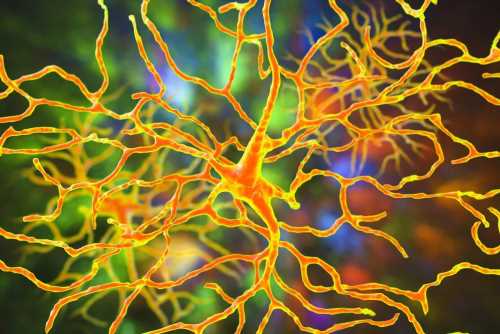
These ROs could be produced in abundance, too, and gave rise to authentic retinal cell progenies. Photoreceptors and RGCs differentiating within these ROs resembled their in vivo counterparts and exhibited all their characteristics, functional and physiological. They also extended axons implying they crosstalked across synapses, with small gaps at their cord’s tips.
First, the team converted hPSC-derived three-dimensional (3D) ROs to a two-dimensional culture system and determined the proportion of different cell classes. On day 80, they dissociated wild-type WA09 hPSC-ROs having a cross-section of neuron subpopulations into a single-cell suspension. They allowed these cells to recover and extend neuronal processes for 20 days before screening with antibodies against neural retina cell fate markers. Finally, the team used high-content image analysis (HCIA) to quantify neural retinal cell populations.
They used monosynaptic retrograde rabies virus (RVdG) tracing for systematically studying de novo synapses among hPSC-retinal neurons. With its G-glycoprotein-deleted, this modified rabies virus, with low neurotoxicity in short-term culture and higher versatility than historically used viral tracers, is widely employed for synaptic tracing studies.
The researchers subjected the 2D cultures to an established RVdG assay on day 10. On day 100, the team examined 2D retinal cell cultures for the presence of starter and traced cells using confocal and high-content widefield imaging. The researchers observed that retinal cells marked by a fluorescent color indicating a rabies infection had infected one across a synapse.
Conclusions
The researchers developed a high-throughput platform to identify and quantify de novo synapses formed among hPSC-derived retinal neurons. They found two major neuron types, photoreceptors, and RGCs, among the traced presynaptic cells.
The study-designed platform for assessing synaptic connections in cultured retinal neurons sets the stage for future cell replacement studies characterizing synaptogenesis. This novel in vitro synaptic tracing method also overcomes the limitations of animal model testing having evolutionary incompatibilities with the synaptic machinery of human xenografts.
Monosynaptic retrograde RVdG tracing emerges as a robust synaptic labeling strategy for studying synaptic connections. This assay incorporates many negative controls to delineate authentic synaptic connections, thus, facilitating the verification of the mechanism governing vision recovery after intraocular transplantation with donor retinal neurons. Additionally, the synapse tracking tools described in this study help address questions of functional synapse formation after allogeneic or xenogeneic transplantation.
Future studies will continue to examine these aspects, which will help better determine the tendency of other retinal neural cell populations derived from hPSC to form new synaptic connections during differentiation. These studies could also characterize the strength of these synapses and their functions.
- Re-formation of synaptic connectivity in dissociated human stem cell-derived retinal organoid cultures, Allison L. Ludwig, Steven J. Mayerl, Yu Gao, Mark Banghart, Cole Bacig, Maria A. Fernandez Zepeda, Xinyu Zhao, David M. Gamm, PNAS 2023, doi: https://doi.org/10.1073/pnas.2213418120
https://www.pnas.org/doi/10.1073/pnas.2213418120
Posted in: Medical Science News | Medical Research News | Medical Condition News | Disease/Infection News
Tags: Animal Model, Antibodies, Assay, Blindness, Cell, Eye, Ganglion, Glaucoma, Glycoprotein, Imaging, in vitro, in vivo, Macular Degeneration, Nerve, Neuron, Neurons, Ophthalmology, Organoids, Rabies, Research, Retinitis Pigmentosa, Stem Cells, Synapse, Virus

Written by
Neha Mathur
Neha is a digital marketing professional based in Gurugram, India. She has a Master’s degree from the University of Rajasthan with a specialization in Biotechnology in 2008. She has experience in pre-clinical research as part of her research project in The Department of Toxicology at the prestigious Central Drug Research Institute (CDRI), Lucknow, India. She also holds a certification in C++ programming.
Source: Read Full Article